
Cataract
 A cataract is a loss of transparency, or clouding, of the normally clear lens of the eye. As one ages, chemical changes occur in the lens that make it less transparent. The loss of transparency may be so mild vision is hardly affected or so severe that no shapes or movements are seen, only light and dark. When the lens gets cloudy enough to obstruct vision to any significant degree, it is called a cataract. Glasses or contact lenses cannot sharpen your vision if a cataract is present.
The most common cause of cataract is aging. Other causes include trauma, medications such as steroids, systemic diseases such as diabetes and prolonged exposure to ultraviolet light. Occasionally, babies are born with a cataract.
Reducing the amount of ultraviolet light exposure by wearing a wide-brim hat and sunglasses may reduce your risk for developing a cataract but once developed there is no cure except to have the cataract surgically removed. Outpatient surgical procedures can remove the cataract through either a small incision (phacoemulsification) or a large incision (extracapsular extraction). The time to have the surgical procedure is when your vision is bad enough that it interferes with your lifestyle.
Cataract surgery is a very successful operation. One and a half million people have this procedure every year and 95% have a successful result. As with any surgical procedure, complications can occur during or after surgery and some are severe enough to limit vision. But in most cases, vision, as well as quality of life, improves.
 Cataract Symptoms
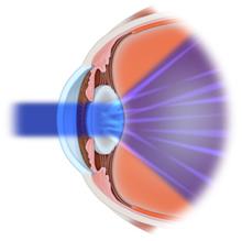
Your eye works a lot like a camera. Light rays focus through your lens on the retina, a layer of light sensitive cells at the back of the eye. Similar to film, the retina allows the image to be "seen" by the brain. But over time the lens can become cloudy and prevent light rays from passing clearly through the lens. This cloudy lens is called a cataract.
The typical symptom of cataract formation is a slow, progressive, and painless decrease in vision. Other changes include: blurring of vision; glare, particularly at night; frequent eyeglass prescription change; a decrease in color intensity; a yellowing of images; and in rare cases, double vision.
Ironically as the lens gets harder, farsighted or hyperopic people experience improved distance vision and are less dependent on glasses. However, nearsighted or myopic people become more nearsighted or myopic, causing distance vision to be worse. Some types of cataracts affect distance vision more than reading vision. Others affect reading vision more than distance vision.


Intraocular Lenses (IOLs)
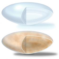
 An intraocular lens (IOL) is a tiny, lightweight, clear plastic disk placed in the eye during cataract surgery. An IOL replaces the focusing power of the eye's natural lens.
The lens of the eye plays an important role in focusing images on the retina. If the lens loses its clarity, as it does when a cataract develops, light rays do not focus clearly and the image one sees is blurry. Glasses or contact lenses cannot sharpen vision if a cataract is present.
The only treatment for a cataract is to remove the lens and implant an IOL. Intraocular lenses have many advantages. Unlike contact lenses, which must be removed, cleaned, and reinserted, the IOL remains in the eye after surgery.
An IOL may be placed either in front of or behind the iris. Behind the iris is the most frequent placement site. They can be hard plastic, soft plastic or soft silicone. Soft, foldable lenses can be inserted through a small incision which shortens recovery time following surgery.
Rapid evolution of IOL designs, materials, and implant techniques have made them a safe and practical way to restore normal vision after cataract surgery.
Phacoemulsification (Phaco)
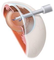
Phacoemulsification is a surgical method used to remove a cataract, which is a clouding of the eye's naturally clear lens. A cloudy lens interferes with light passing through to the retina, the light-sensing layer of cells at the back of the eye. Having a cataract can be compared to looking at the world through a foggy window.
In phacoemulsification, an ultrasonic oscillating probe is inserted into the eye. The probe breaks up the center of the lens. The fragments are suctioned from the eye at the same time. A small incision that often does not require sutures to close can be used since the cataract is removed in tiny pieces. Most of the lens capsule is left behind and a foldable intraocular lens implant, or IOL, is placed permanently inside to help focus light onto the retina. Vision returns quickly and one can resume normal activities within a short period of time.
YAG Capsulotomy (Laser)
When your cataract was removed, your eye's lens was replaced with an IOL. This clear lens is placed in the posterior capsule, which held the old lens. You could see again because the IOL allowed light to reach your retina. The posterior capsule is a thin, clear film, much like a piece of cellophane. Months or even years after cataract surgery, the posterior capsule can become cloudy. This may block light from reaching the retina. This is sometimes called "after or secondary cataract," but it is not a new cataract. If this cloudiness occurs, a laser treatment called, YAG capsulotomy, is performed. Your doctor uses a YAG laser, which delivers tiny, rapid bursts of energy. The laser passes through the front of the eye and the IOL without harming them. When the laser reaches the posterior capsule, it makes a tiny opening. Light can then enter the eye again. Enough of the posterior capsule is left to hold the IOL in place.
The procedure is quick and painless. There are no needles or stitches. Your eye is numbed with drops and your pupil is dilated with drops. Then you rest your chin on a stand in front of the laser. You may see flashes of light and hear a faint clicking sound, but you should not feel any pain. You should begin to see better within a few hours. Your doctor may check your eye pressure later that day or the next. You can most often return to your normal routine right away. Your vision will most likely be fully restored soon after treatment.
An intraocular lens (IOL) is a tiny, lightweight, clear plastic disk placed in the eye during cataract surgery. An IOL replaces the focusing power of the eye's natural lens.
The lens of the eye plays an important role in focusing images on the retina. If the lens loses its clarity, as it does when a cataract develops, light rays do not focus clearly and the image one sees is blurry. Glasses or contact lenses cannot sharpen vision if a cataract is present.
The only treatment for a cataract is to remove the lens and implant an IOL. Intraocular lenses have many advantages. Unlike contact lenses, which must be removed, cleaned, and reinserted, the IOL remains in the eye after surgery.
An IOL may be placed either in front of or behind the iris. Behind the iris is the most frequent placement site. They can be hard plastic, soft plastic or soft silicone. Soft, foldable lenses can be inserted through a small incision which shortens recovery time following surgery.
Rapid evolution of IOL designs, materials, and implant techniques have made them a safe and practical way to restore normal vision after cataract surgery.
Phacoemulsification (Phaco)
Phacoemulsification is a surgical method used to remove a cataract, which is a clouding of the eye's naturally clear lens. A cloudy lens interferes with light passing through to the retina, the light-sensing layer of cells at the back of the eye. Having a cataract can be compared to looking at the world through a foggy window.
In phacoemulsification, an ultrasonic oscillating probe is inserted into the eye. The probe breaks up the center of the lens. The fragments are suctioned from the eye at the same time. A small incision that often does not require sutures to close can be used since the cataract is removed in tiny pieces. Most of the lens capsule is left behind and a foldable intraocular lens implant, or IOL, is placed permanently inside to help focus light onto the retina. Vision returns quickly and one can resume normal activities within a short period of time.
YAG Capsulotomy (Laser)
When your cataract was removed, your eye's lens was replaced with an IOL. This clear lens is placed in the posterior capsule, which held the old lens. You could see again because the IOL allowed light to reach your retina. The posterior capsule is a thin, clear film, much like a piece of cellophane. Months or even years after cataract surgery, the posterior capsule can become cloudy. This may block light from reaching the retina. This is sometimes called "after or secondary cataract," but it is not a new cataract. If this cloudiness occurs, a laser treatment called, YAG capsulotomy, is performed. Your doctor uses a YAG laser, which delivers tiny, rapid bursts of energy. The laser passes through the front of the eye and the IOL without harming them. When the laser reaches the posterior capsule, it makes a tiny opening. Light can then enter the eye again. Enough of the posterior capsule is left to hold the IOL in place.
The procedure is quick and painless. There are no needles or stitches. Your eye is numbed with drops and your pupil is dilated with drops. Then you rest your chin on a stand in front of the laser. You may see flashes of light and hear a faint clicking sound, but you should not feel any pain. You should begin to see better within a few hours. Your doctor may check your eye pressure later that day or the next. You can most often return to your normal routine right away. Your vision will most likely be fully restored soon after treatment.